Skip to content

Teach Human Reproduction in eight steps.

For each idea: show it, ask before you tell, then say one line.

Gather paper and a pencil.

Common misconceptions & worked visual for this chapter · 15 practice questions with answers

How to teach it

Reproductive organs and cells

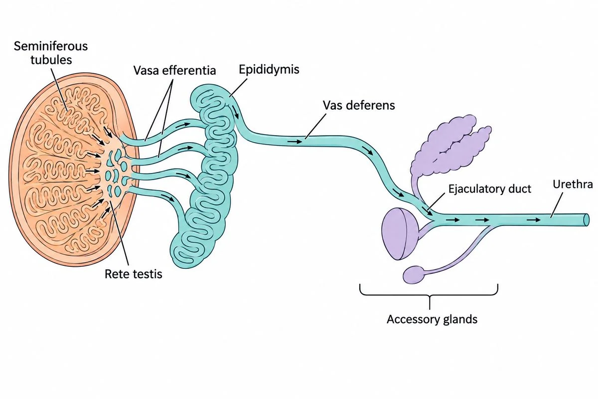
Labelled sperm route with directional arrows, an enlarged testis inset and accessory gland branches.

1. Male reproductive system

Testes make sperm; ducts carry them; glands add fluid.

Show
Look at the diagram. Trace the labelled sperm route from seminiferous tubules to urethra. Find where the accessory gland branches add fluid.
Ask
What will those glands add to the passing sperm?
Say
Glands add fluid, making semen.
Used in
Semen testing helps investigate infertility.
If your child says…“The testes make all the semen.”
Ask
Why do gland branches join the sperm route?
What’s true
Accessory glands supply most seminal fluid.
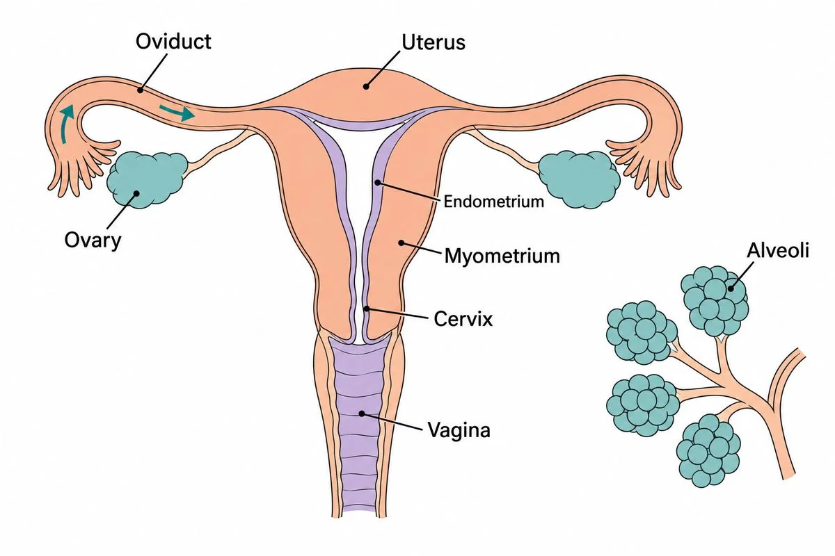
Female reproductive organs showing uterine layers, with a separate mammary alveoli inset.

2. Female reproductive system

Ovaries supply oocytes; the uterus supports pregnancy; mammary glands make milk.

Show
In the diagram, trace the released oocyte from the ovary into the oviduct, towards the uterus. Compare lining and muscle. Find the alveoli in the separate mammary inset.
Ask
Where will an embryo attach after travelling along this route?
Say
The uterine lining receives the embryo. The myometrium supplies birth contractions. Mammary alveoli make milk.
Used in
Ultrasound helps locate a pregnancy within the uterus.
If your child says…“The inner lining pushes the baby out.”
Ask
Which layer is muscle?
What’s true
The muscle layer supplies the powerful contractions.
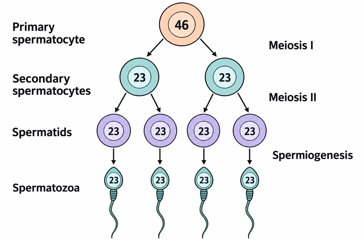
Two divisions produce four spermatids, followed by their change into sperm.

3. Spermatogenesis

A primary spermatocyte divides twice to produce four future sperm.

Show
Look at the diagram. Follow both divisions, comparing cell numbers and chromosome counts. Trace the final change from round spermatids to tailed sperm.
Ask
After both divisions shown, how will cell number and chromosome number per cell change?
Say
The first division halves the chromosome number. The second produces four spermatids. Spermiogenesis reshapes them into sperm without another division.
Used in
Examining sperm shape helps investigate fertility problems.
If your child says…“One starting cell makes only two sperm.”
Ask
What happens when both first-division cells divide again?
What’s true
Meiosis includes another division after the first.

From ovulation to pregnancy

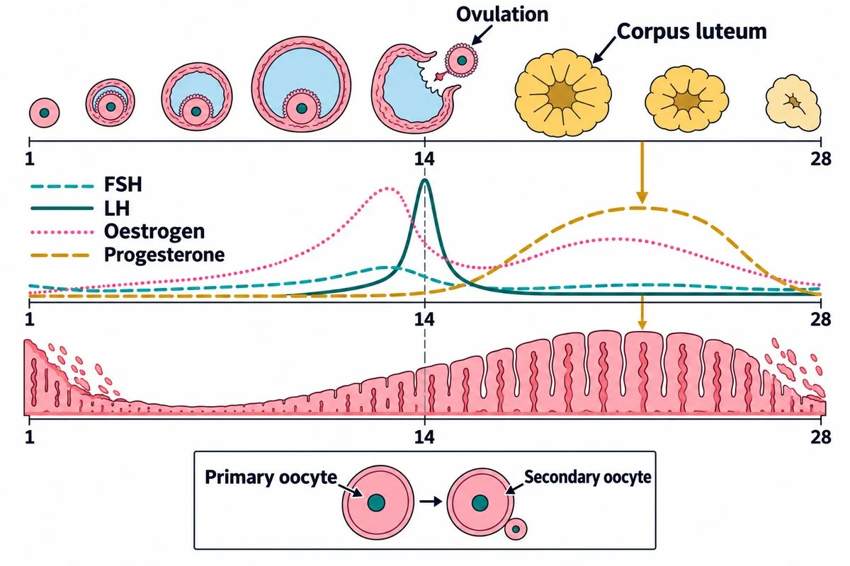
Aligned ovarian changes, hormone curves and uterine lining across an illustrative menstrual cycle.

4. Oogenesis and menstrual cycle

Ovarian hormones coordinate oocyte development with changes in the uterine lining.

Show
Look at the diagram’s unequal oocyte division. Align the LH peak with ovulation, then follow the empty follicle becoming the corpus luteum. Compare hormone curves with the lining below.
Ask
How will changing ovarian hormones affect the lining while the oocyte develops elsewhere?
Say
Oestrogen rebuilds the lining, and progesterone maintains it after ovulation. When these hormones fall, the lining sheds.
Used in
Period records help describe cycle changes during health consultations.
If your child says…“Rising hormones start a period.”
Ask
What happens when the lining loses its hormonal support?
What’s true
Menstruation follows a fall in ovarian hormones.
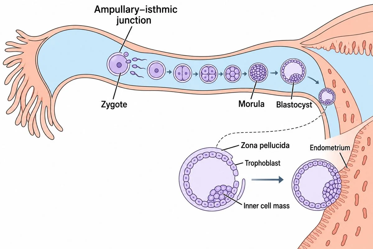
Fertilisation and cleavage along an oviduct, followed by blastocyst attachment inside the uterus.

5. Fertilisation and implantation

Sperm and egg unite; the developing embryo attaches inside the uterus.

Show
Follow the diagram from fertilisation to blastocyst. Find its outer layer beside the uterine lining.
Ask
How will the blastocyst attach to the uterine lining?
Say
The trophoblast attaches to the lining.
Used in
IVF allows embryos to develop before transfer into the uterus.
If your child says…“The outer layer becomes the whole baby.”
Ask
Which cluster lies inside the attaching outer layer?
What’s true
The embryo develops from the inner cell mass.
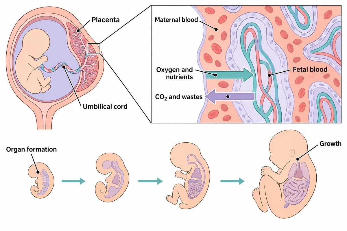
Placental exchange across separate blood supplies, above stages of organ formation and growth.

6. Pregnancy and embryonic development

The placenta supports pregnancy while the embryo’s organs gradually develop.

Show
Look at the diagram’s placenta and umbilical connection. Follow exchange arrows across the tissue separating the blood supplies. Compare early organ formation with later growth below.
Ask
What will oxygen cross to reach fetal blood when these blood supplies stay separate?
Say
Oxygen and nutrients cross placental tissue into fetal blood, while wastes pass the other way. Organs form gradually, then grow and mature.
Used in
Pregnancy tests detect hCG, helping identify pregnancy.
If your child says…“All the organs are ready at implantation.”
Ask
What changes between the early and later developmental outlines?
What’s true
Organ formation continues after implantation.

Birth and feeding

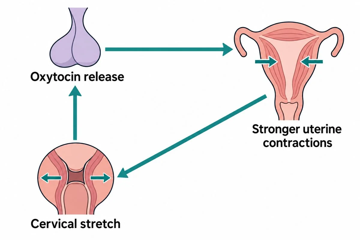
Labelled clockwise loop linking cervical stretch, oxytocin release and stronger uterine contractions.

7. Parturition

Stretch and oxytocin reinforce uterine contractions until birth.

Show
Look at the diagram. Follow the arrows from cervical stretch to oxytocin release, then stronger uterine contractions. Follow the returning arrow to more cervical stretch.
Ask
What will another turn around this loop do to the contractions?
Say
Stretch triggers oxytocin release, strengthening contractions and causing more stretch.
Used in
Monitoring contractions helps maternity staff follow labour’s progress.
If your child says…“Each contraction makes the next one weaker.”
Ask
Does more stretch trigger less oxytocin or more?
What’s true
Positive feedback strengthens the response.
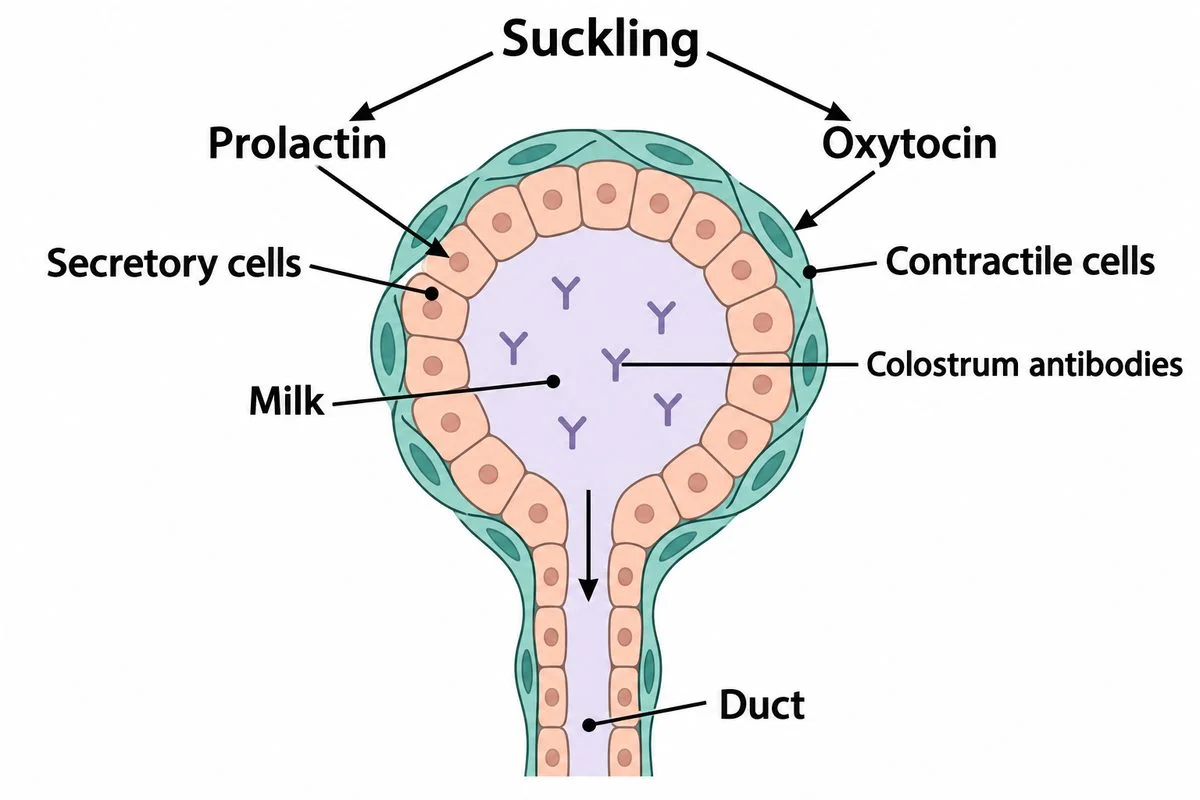
Two hormone pathways control milk production and release from a mammary alveolus.

8. Lactation

Hormones control milk production and release; colostrum supplies protective antibodies.

Show
Look at the diagram’s two paths from suckling. Follow prolactin to the secretory cells and oxytocin to the surrounding contractile cells. Trace the milk’s route into the duct.
Ask
How will suckling help both make fresh milk and release milk already present?
Say
Prolactin helps the lining cells make milk. Oxytocin makes surrounding cells squeeze stored milk into the duct.
Used in
Colostrum supplies maternal antibodies, helping protect newborns against infection.
If your child says…“Oxytocin makes the milk.”
Ask
Which labelled cells can squeeze the sac?
What’s true
Oxytocin acts on the surrounding muscle cells to squeeze milk out.

A real problem

IVF can help when Fallopian tubes are blocked.

Blocked Fallopian tubes can cause infertility.

The technique allowed embryos to develop outside the body before transfer into the uterus.

The first successful human birth using IVF occurred in 1978.

Nobel PrizeScience Museum Group

Clear Petri dish beside a microscope on a laboratory bench.

Concept Lab · Common misconception

The tempting mistake in Human Reproduction

Open standalone misconception page →

Where does fertilisation usually happen?

Wrong

Fertilisation usually occurs in the uterus because that is where the baby develops.

Why it feels right. I picture every stage happening in the same place.

Right

Fertilisation usually occurs in a uterine tube. The early embryo later implants in the uterus.

Worked example

Sequence
Egg enters uterine tube→Fertilisation usually in tube→Embryo travels→Implantation in uterus
Fertilisation and implantation occur at different locations.
  1. Trace an ovulated egg into the uterine tube.
  2. After fertilisation the early embryo travels towards the uterus, where implantation can occur.

Ask your child eight quick questions.

One per step. The answer and the reason are given.

  1. Where does most seminal fluid come from?

    The accessory glands. Testes produce sperm; accessory glands contribute most of the fluid.

  2. Which uterine layer contracts strongly during childbirth?

    The myometrium. The myometrium is muscle; the endometrium receives the embryo.

  3. How many spermatids result from one primary spermatocyte?

    Four. Both cells formed by the first division divide again.

  4. What change in ovarian hormones triggers menstruation?

    Their levels fall. The lining sheds when progesterone and oestrogen no longer maintain it.

  5. Which part of the blastocyst develops into the embryo?

    The inner cell mass. The trophoblast attaches outside; the embryo develops from the inner cluster.

  6. When do organs develop: before implantation or progressively afterwards?

    Progressively afterwards. An implanting blastocyst does not yet have complete organs.

  7. What happens to uterine contractions when oxytocin increases during labour?

    They become stronger. Oxytocin strengthens contractions, increasing stretch and encouraging further oxytocin release.

  8. Which hormone causes already-made milk to flow out?

    Oxytocin. Oxytocin contracts surrounding muscle cells; prolactin supports making milk.
15 more questions with answers