Skip to content

Teach Excretory Products and Their Elimination in eight steps.

For each idea: show it, ask before you tell, then say one line.

Gather paper and a pencil.

Common misconceptions & worked visual for this chapter · 15 practice questions with answers

How to teach it

Wastes and their routes

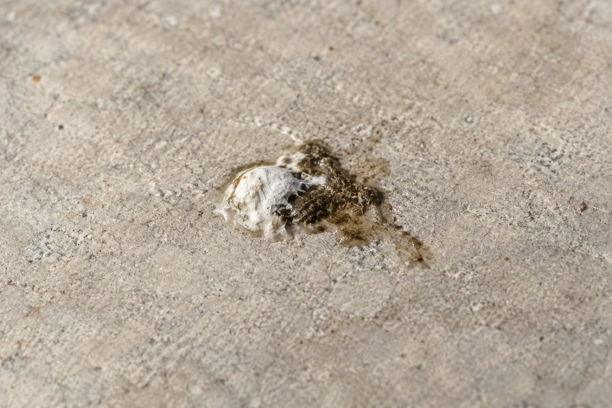
A bird dropping with whitish paste and darker faeces on paving.

1. Ammonotelism, ureotelism and uricotelism

Removing nitrogen waste as ammonia, urea or uric acid.

Show
Find a bird dropping outdoors without touching it. Point out its whitish paste and darker faeces.
Ask
Would removing this waste in watery urine require more water?
Say
Yes; birds conserve water with uric acid.
Used in
Earthworm nephridia remove wastes and regulate water and salts, keeping internal fluids balanced.
If your child says…“Ammonia needs the least water.”
Ask
Which needs more water for removal: dissolved ammonia or whitish uric acid paste?
What’s true
Ammonia is more toxic and requires more water for removal than uric acid.
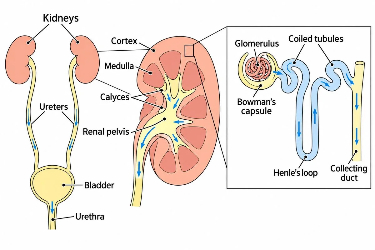
Labelled urinary organs, kidney cutaway and enlarged nephron showing the route of urine.

2. Human excretory system

Kidneys make urine; connected passages carry, store and release it.

Show
Look at the diagram. Trace from the capsule through both coils and the loop to the collecting duct. Continue through calyces, renal pelvis and ureter to the bladder and urethra.
Ask
Where will fluid leaving the collecting duct go?
Say
Urine follows calyces, pelvis and ureter to the bladder, then exits through the urethra.
Used in
Tracing urine passages helps explain where a kidney stone can obstruct drainage.
If your child says…“The glomerulus alone is the renal corpuscle.”
Ask
Does the diagram include only the capillary knot, or its surrounding cup too?
What’s true
The renal corpuscle includes both the blood capillaries and the capsule receiving their filtrate.

How kidneys form and adjust urine

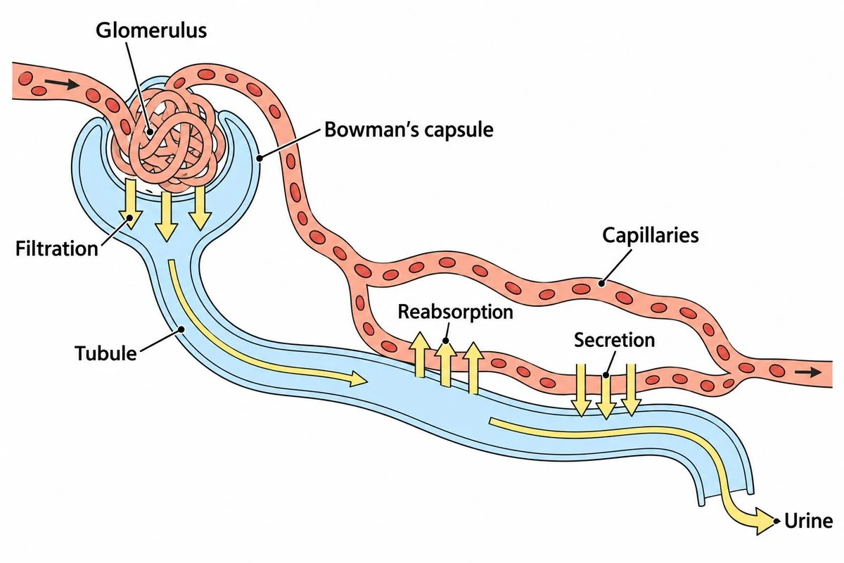
Nephron diagram showing filtration, reabsorption and secretion between blood and tubule.

3. Urine formation

Filtering blood, reclaiming useful substances and adding selected wastes.

Show
Find the glomerulus inside the diagram’s capsule. Follow the arrows between blood and tubule.
Ask
Must glucose entering this capsule be lost in urine?
Say
No. Filtration lets glucose enter the capsule while blood cells and most proteins remain in blood. Reabsorption normally returns this glucose to blood.
Used in
Reabsorption keeps filtered nutrients available for the body to use.
If your child says…“Moving glucose back into blood is secretion.”
Ask
Is this glucose returning to blood or entering the tubule?
What’s true
Reabsorption returns substances to blood; secretion moves substances into tubular fluid.
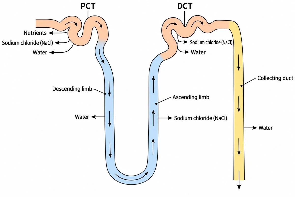
Tubule sections with different arrows for recovering nutrients, salts and water.

4. Function of the tubules

Different tubule sections recover different substances from filtered fluid.

Show
Trace the diagram from PCT through both limbs to DCT and collecting duct. Compare outward arrows.
Ask
How will the ascending limb’s fluid concentration change when salts leave but water stays?
Say
The PCT recovers nutrients, salts and water, while the descending limb lets water leave. The ascending limb releases salts but retains water, diluting its fluid. The DCT adjusts salts and water, while the collecting duct adjusts water recovery.
Used in
DCT and collecting-duct adjustments help keep blood water, salts and acidity balanced.
If your child says…“Fluid becomes more concentrated as it rises.”
Ask
If salt leaves but water stays, what happens to the salt concentration?
What’s true
Removing salts while retaining water makes the ascending fluid more dilute.
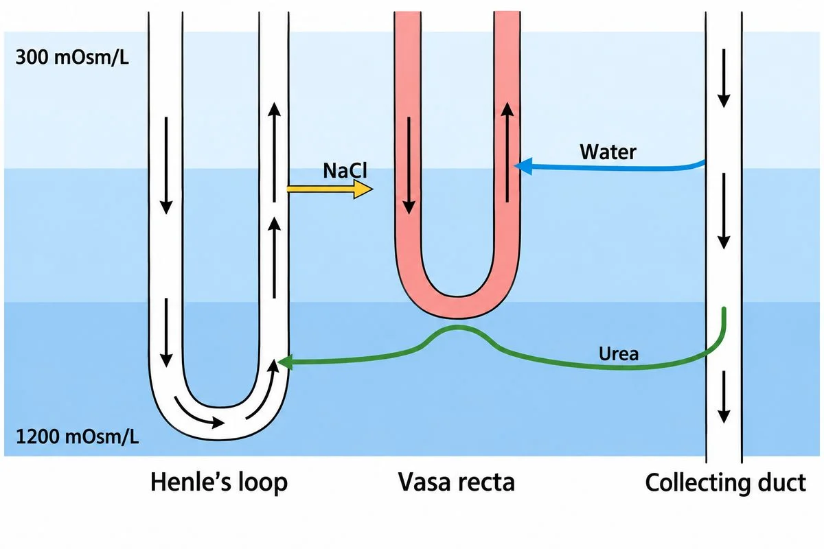
Opposite flows preserve concentrated surroundings that draw water from the collecting duct.

5. Mechanism of concentration of the filtrate

Concentrated surroundings draw water from a collecting duct that permits passage.

Show
Compare the diagram’s concentrations. Follow the opposite flows.
Ask
How can these surroundings help recover water?
Say
Salt and urea concentrate the medulla; vasa recta exchange preserves this gradient. Water leaves by osmosis and returns to blood.
Used in
Concentrated urine lets kidneys remove wastes while conserving water.
If your child says…“The surroundings get less concentrated deeper down.”
Ask
Which end of the diagram shows the higher surrounding concentration?
What’s true
Fluid around the tubules becomes more concentrated towards the inner medulla.
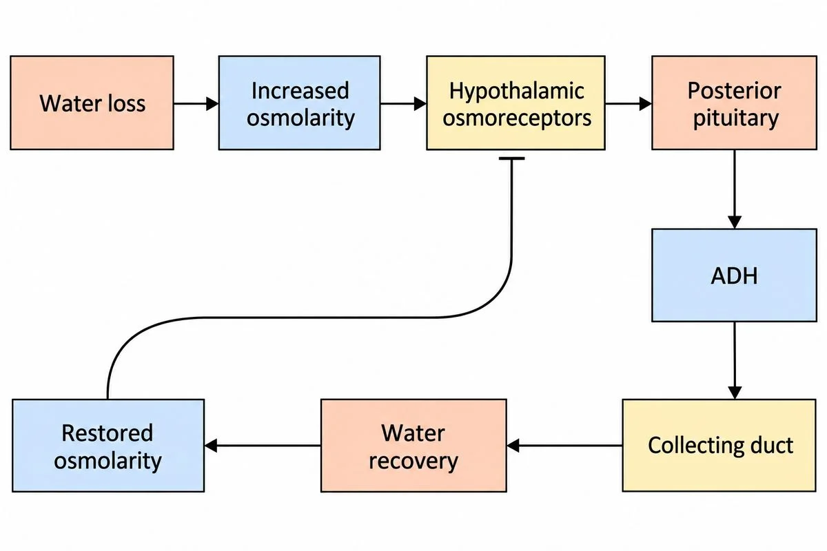
Feedback diagram linking water loss, ADH, kidney water recovery and reduced stimulation.

6. Regulation of kidney function

ADH adjusts water recovery through feedback.

Show
Follow the diagram from water loss through osmoreceptors to ADH. Trace the return line.
Ask
How would the collecting duct conserve water after the water loss shown here?
Say
Osmoreceptors detect more concentrated blood, prompting ADH release from the posterior pituitary. ADH increases water recovery by the collecting duct. Restored water balance reduces the signal for further ADH release.
Used in
During water shortage, ADH reduces water lost in urine.
If your child says…“ADH makes us pass more urine.”
Ask
If more water returns to blood, how much remains for urine?
What’s true
ADH increases water recovery in later tubule sections, leaving less water in urine.

Passing urine and replacing lost kidney function

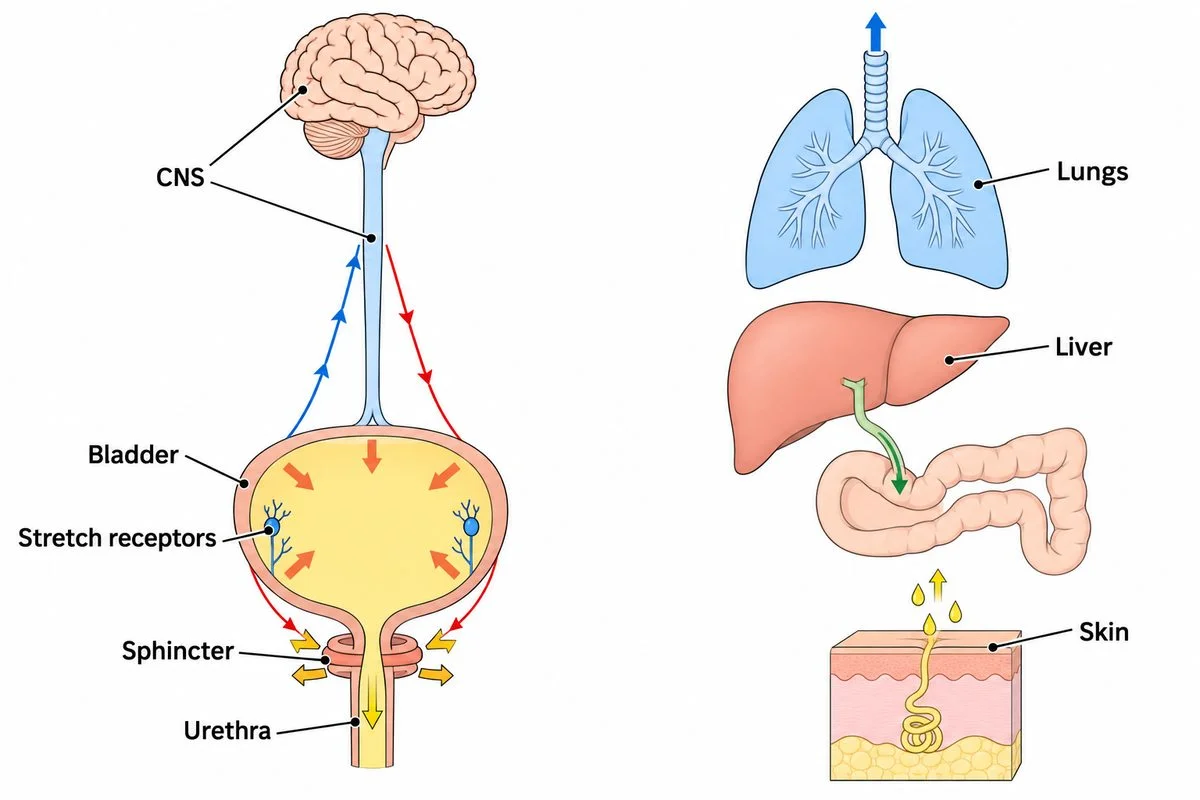
Bladder nerve pathway beside lungs, liver and skin showing additional waste routes.

7. Micturition and role of other organs in excretion

Nerves coordinate urination; lungs, liver and skin also remove wastes.

Show
Trace bladder signals through the diagram’s CNS. Find the other excretory organs.
Ask
How will these signals empty the bladder?
Say
Nerves coordinate bladder contraction and sphincter relaxation; the external sphincter has voluntary control. Lungs excrete carbon dioxide and water; the liver excretes through bile. Sweat removes water, salts and some urea.
Used in
Sweat evaporation cools us while sweat also carries away small amounts of waste.
If your child says…“A full bladder empties without nerves.”
Ask
How does information about a filling bladder reach your brain?
What’s true
Bladder stretch receptors signal the CNS, which sends messages coordinating emptying.
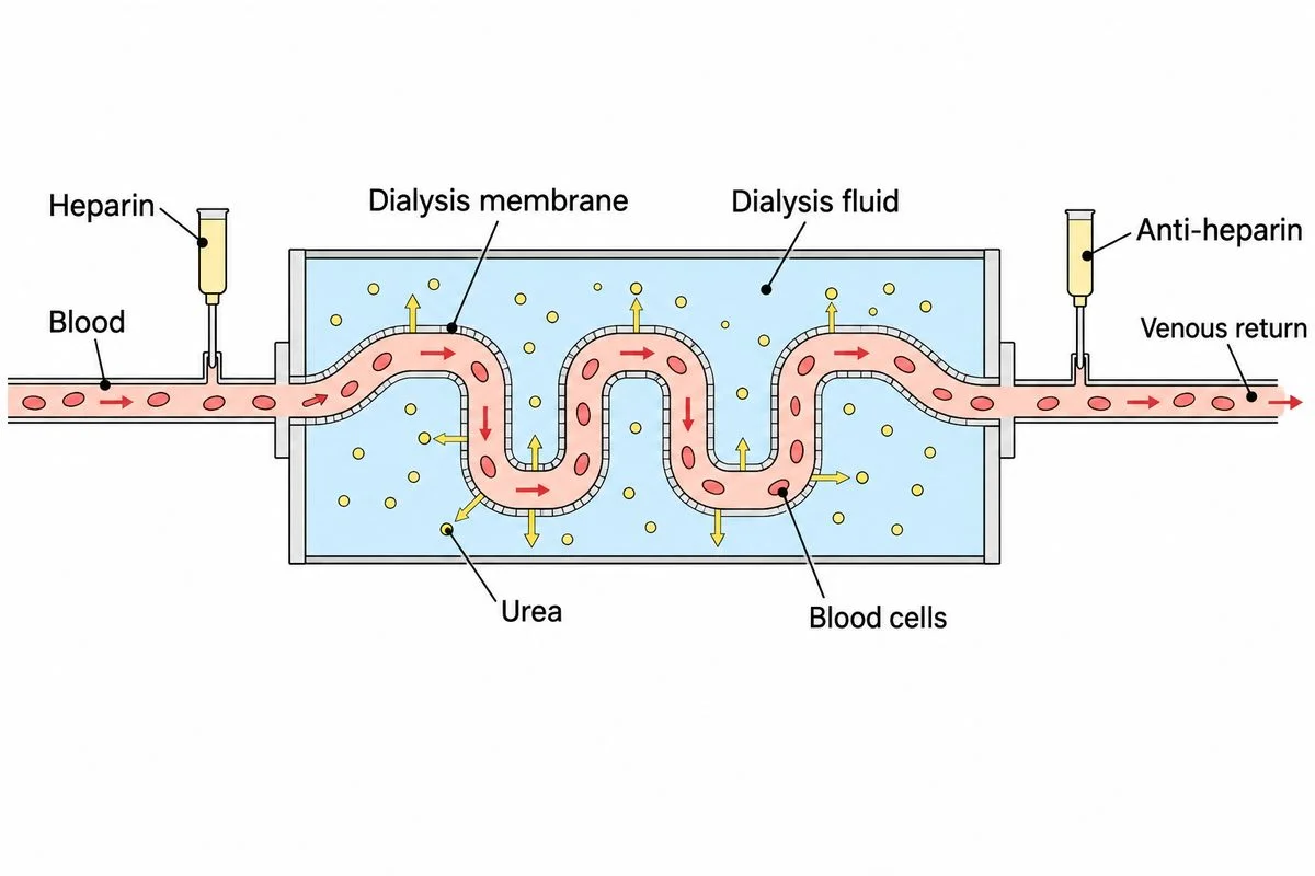
Dialysis diagram showing blood flow, drug placement and urea crossing into dialysis fluid.

8. Disorders of the excretory system

Kidney damage can leave wastes in blood; treatment replaces lost function.

Show
Copy the tube inside a box: blood inside, dialysis fluid outside. Label heparin before the dialyser and anti-heparin before venous return.
Ask
Where will urea move if surrounding fluid contains none?
Say
Heparin prevents clotting before blood enters the dialyser. Urea crosses into dialysis fluid; blood cells stay inside. Anti-heparin restores normal clotting before blood returns through a vein.
Used in
Haemodialysis removes urea from blood when failing kidneys cannot clear it adequately.
If your child says…“The dialysis fluid must be full of waste.”
Ask
Which side starts with more urea: the blood or the surrounding fluid?
What’s true
Incoming dialysis fluid lacks nitrogenous wastes, allowing their movement out of blood.

A real problem

A transplanted kidney can take over blood filtration.

CMC Vellore performed its pioneering kidney transplant in 1971.

Kidney transplantation treats advanced, permanent kidney failure.

The transplanted kidney takes over filtering the blood.

CMC VelloreHarvard HealthNIDDK

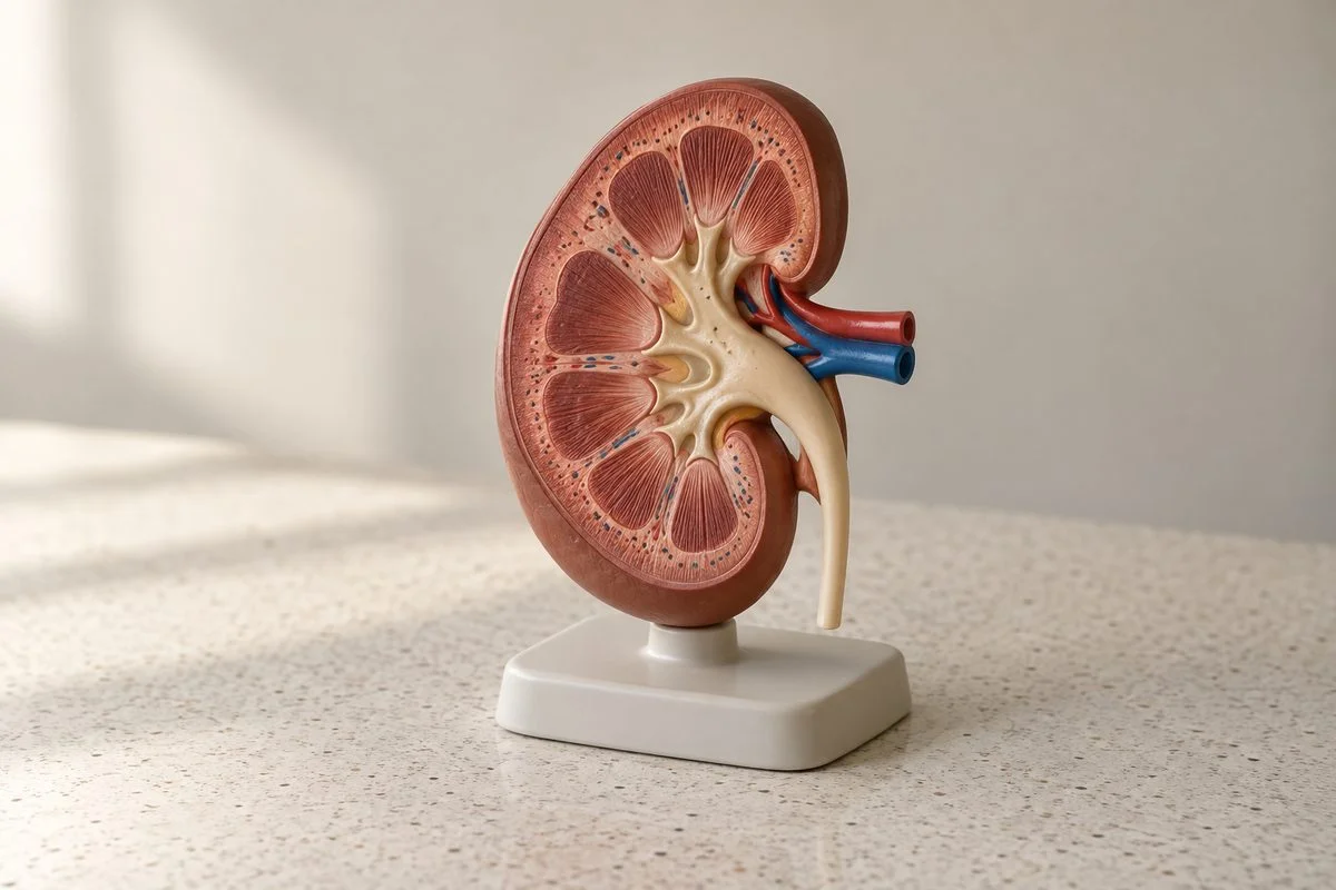
A kidney anatomy model on a consultation desk.

Concept Lab · Common misconception

The tempting mistake in Excretory Products and Their Elimination

Open standalone misconception page →

Is everything filtered by the kidney lost in urine?

Wrong

All filtered glucose ends up in urine.

Why it feels right. Filtration sounds like a one-way removal step.

Right

The tubules reabsorb useful filtered substances.

Worked example

Glucose path
Blood→Filtrate→Reabsorbed in tubule→Blood
Filtered glucose enters the tubule first. Under normal conditions nearly all is transported back toward the blood.
  1. Trace glucose from the blood into the filtrate.
  2. In normal conditions most or all filtered glucose is taken back into the blood along the tubules.

Ask your child eight quick questions.

One per step. The answer and the reason are given.

  1. Which nitrogenous waste lets birds lose very little water?

    Uric acid. It can leave as paste with little water.

  2. What surrounds the glomerulus to complete the renal corpuscle?

    Bowman’s capsule. The capillary knot and surrounding capsule together form this structure.

  3. What process returns filtered glucose to blood?

    Reabsorption. Secretion moves substances towards tubular fluid, in the opposite direction.

  4. What leaves the ascending limb while water stays inside?

    Salts. Salt loss without water loss dilutes the fluid.

  5. Where are the surroundings more concentrated: cortex or inner medulla?

    Inner medulla. The inward rise in concentration supports water recovery from the collecting duct.

  6. With other conditions unchanged, what happens to urine volume when ADH increases?

    It decreases. More water returns to blood from the later tubule sections.

  7. Where do bladder stretch receptors send their signals?

    The central nervous system. Nerve messages coordinate bladder contraction and opening of the outlet.

  8. Which way does urea move when incoming dialysis fluid contains none?

    From blood into dialysis fluid. Urea crosses towards the side with a lower urea concentration.

15 more questions with answers, 6 with pictures