Skip to content

Teach Locomotion and Movement in eight steps.

For each idea: show it, ask before you tell, then say one line.

Gather chalk.

Common misconceptions & worked visual for this chapter · 15 practice questions with answers

How to teach it

How muscles produce movement

A chalk starting line on a clear floor.

1. Movement and locomotion

Movement becomes locomotion when the whole organism changes place.

Show
Mark a starting line. After the prediction, your child marches there, then walks forwards.
Ask
Which action will take you away from the line?
Say
Walking changes your place; marching there moves parts of you.
Used in
Skeletal muscles usually follow conscious commands, allowing deliberate movements such as writing.
If your child says…“Every movement is locomotion.”
Ask
When you marched on the spot, did your place change?
What’s true
Moving a part does not necessarily move the whole organism elsewhere.
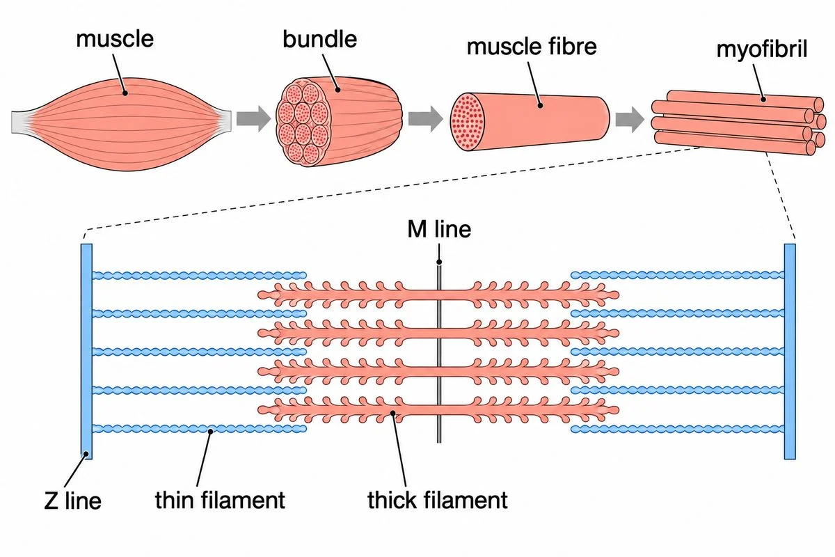
Muscle organisation and an enlarged sarcomere with labelled filaments, Z lines and M line.

2. Skeletal muscle and sarcomere

Muscle cells contain myofibrils divided into repeating contraction units.

Show
Look at the diagram. Follow the muscle bundles to individual fibres, then their myofibrils. Find thin filaments extending inward from Z lines, with the M line midway between them.
Ask
Which boundaries will move together when this unit contracts?
Say
These boundaries are Z lines; the unit between them is a sarcomere. Each muscle fibre contains many myofibrils.
Used in
Sarcomeres shorten together so skeletal muscles can lift a schoolbag.
If your child says…“A sarcomere runs from one middle line to the next.”
Ask
Which lines anchor the thin filaments at the unit's ends?
What’s true
Z lines bound the sarcomere; its M line lies centrally.
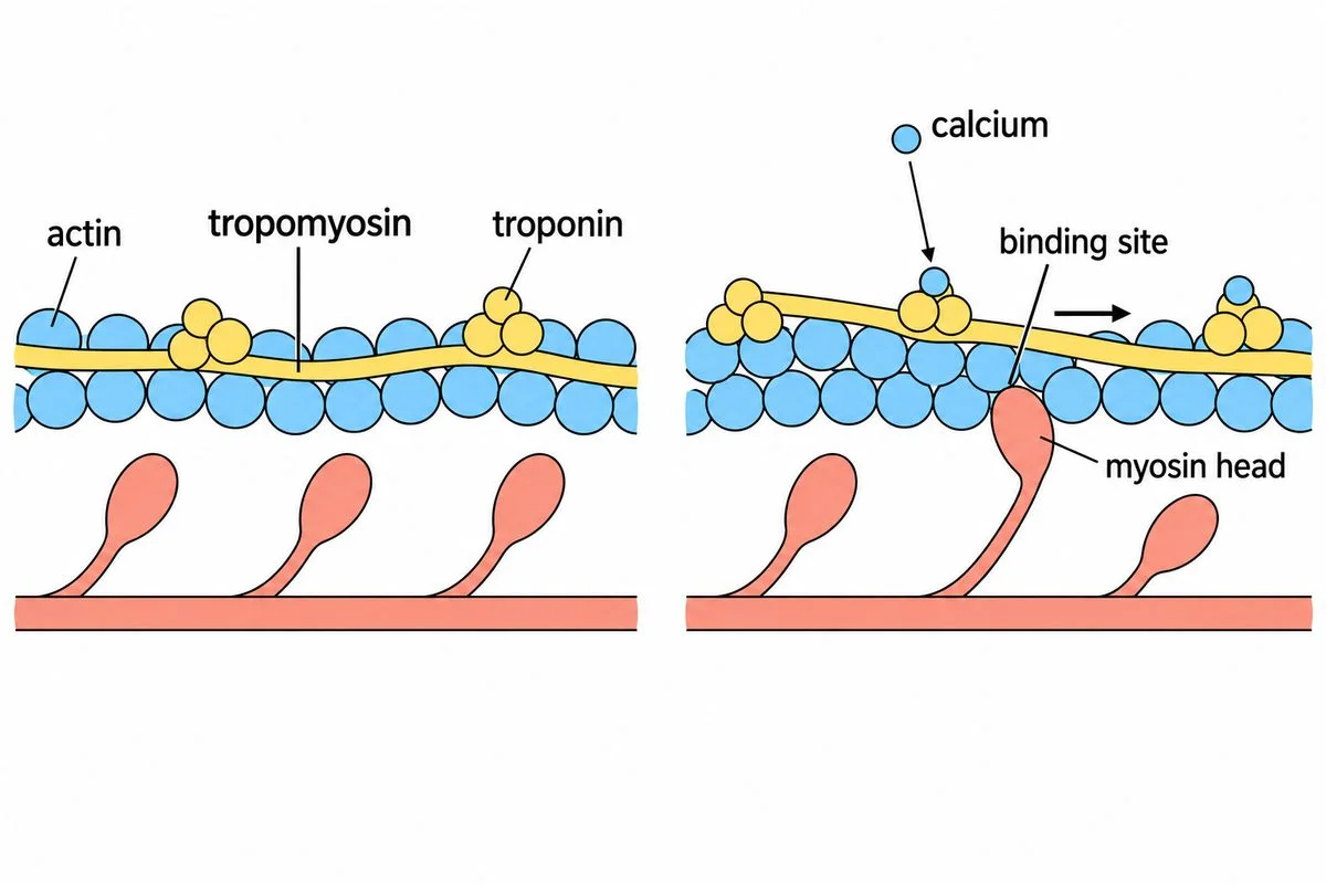
Paired filament diagrams show calcium binding to troponin and exposing actin's attachment sites.

3. Contractile proteins

Myosin grips actin when control proteins expose attachment sites on actin.

Show
Look at the diagram's left panel. Find tropomyosin covering actin's binding sites, with troponin alongside. After predicting, follow calcium to troponin in the right panel.
Ask
When calcium binds troponin, what will change about these myosin heads' ability to attach?
Say
At rest, troponin and tropomyosin keep actin's binding sites covered. Calcium binds troponin, shifting tropomyosin so myosin heads can attach.
Used in
These control proteins regulate muscle contraction, allowing muscles to respond to signals.
If your child says…“Tropomyosin does everything; troponin has no job.”
Ask
Which protein binds calcium before tropomyosin shifts?
What’s true
Troponin responds to calcium and helps control whether myosin can attach.
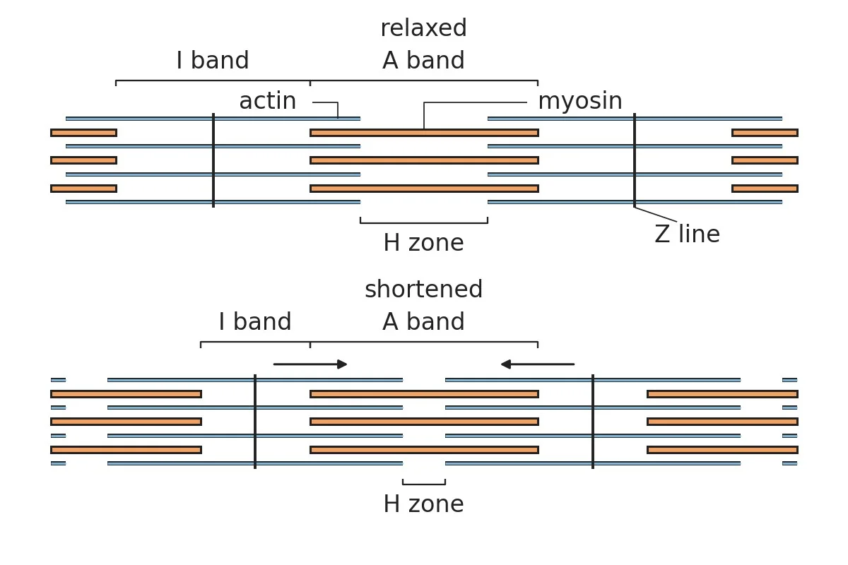
Aligned sarcomeres show increased overlap, closer Z lines and an unchanged A band.

4. Sliding filament theory

Filaments slide past one another, shortening the sarcomere.

Show
Look at the diagram's upper sarcomere. Find its Z lines, actin and myosin. After predicting, compare the lower sarcomere's Z lines and labelled bands.
Ask
If actin slides farther past myosin, what will happen to the distance between Z lines?
Say
Actin slides past myosin, bringing the Z lines closer without shortening either filament. The I band and H zone narrow. The A band keeps its width.
Used in
Repeated filament sliding lets chewing muscles move the jaw through a meal.
If your child says…“The dark A band gets shorter.”
Ask
Which bracket spans the same length of myosin in both diagrams?
What’s true
The A band spans the thick filaments, whose length remains unchanged.

Bones and joints

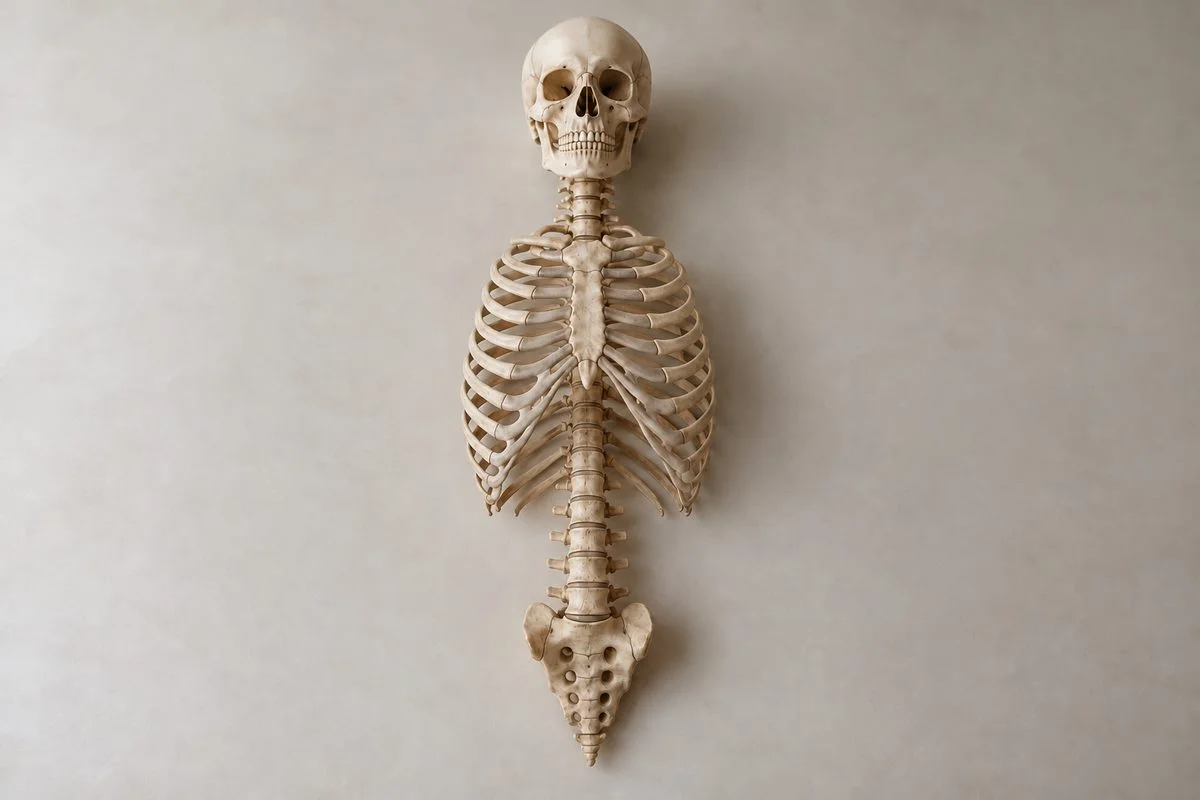
An articulated specimen showing the skull, backbone, sternum and ribs.

5. Axial skeleton

The skull, backbone, breastbone and ribs form the central skeleton.

Show
Gently locate your skull, breastbone, ribs and accessible backbone bumps. Find their arrangement in the photograph.
Ask
Where do you predict your wrist will lie relative to your central skeleton?
Say
Your wrist lies away from these central bones, which form the axial skeleton.
Used in
The rib cage supports the chest while shielding organs needed for breathing and circulation.
If your child says…“All 206 bones belong to the axial skeleton.”
Ask
Is your wrist part of the central framework you just located?
What’s true
The adult axial skeleton contains 80 bones; limbs and girdles belong to another division.
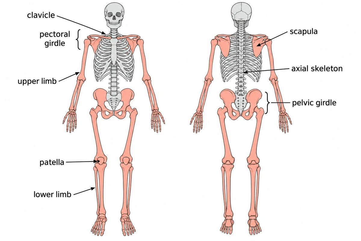
Front and back skeleton diagrams highlight limb bones, supporting girdles and the patella.

6. Appendicular skeleton

Limb bones and their supporting girdles form this skeletal division.

Show
Look at the diagram's front and back views. Trace each limb to its highlighted girdle. Find the patella at the knee's front.
Ask
When you raise your arm, which highlighted girdle will support it?
Say
Limb bones and their supporting girdles make up the appendicular skeleton. The pectoral girdle supports upper limbs, and the pelvic girdle supports lower limbs. The patella belongs at the knee.
Used in
The pelvic girdle connects the lower limbs to the trunk, supporting standing and walking.
If your child says…“The patella belongs at the elbow.”
Ask
Which labelled bone lies at the front of the knee?
What’s true
The patella sits within the thigh muscle's tendon at the front of the knee.
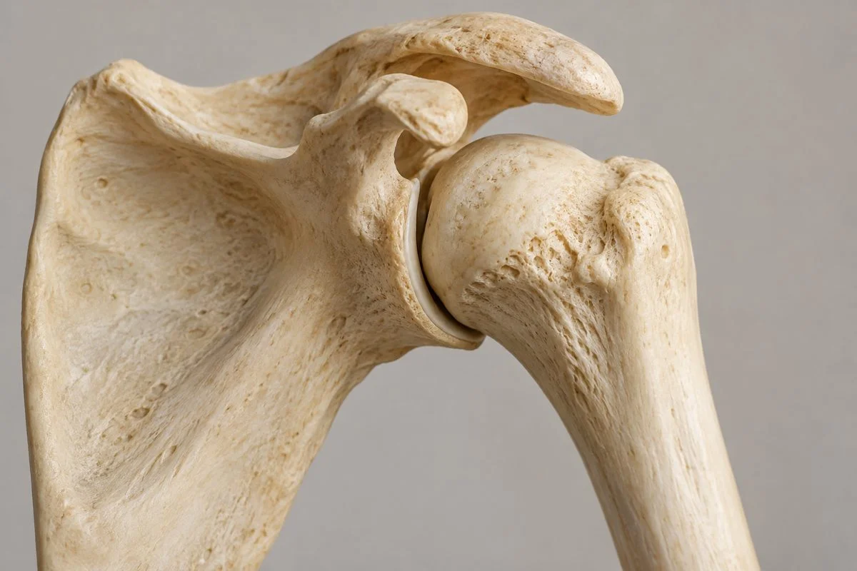
An anatomical shoulder specimen showing its rounded head and socket.

7. Joints

Joint structure determines the directions and extent of bone movement.

Show
After predicting, keep your upper arm still and gently bend your elbow. Then make a small, comfortable shoulder circle.
Ask
Which joint will allow movement in more directions?
Say
Your elbow mainly bends and straightens; your shoulder also turns.
Used in
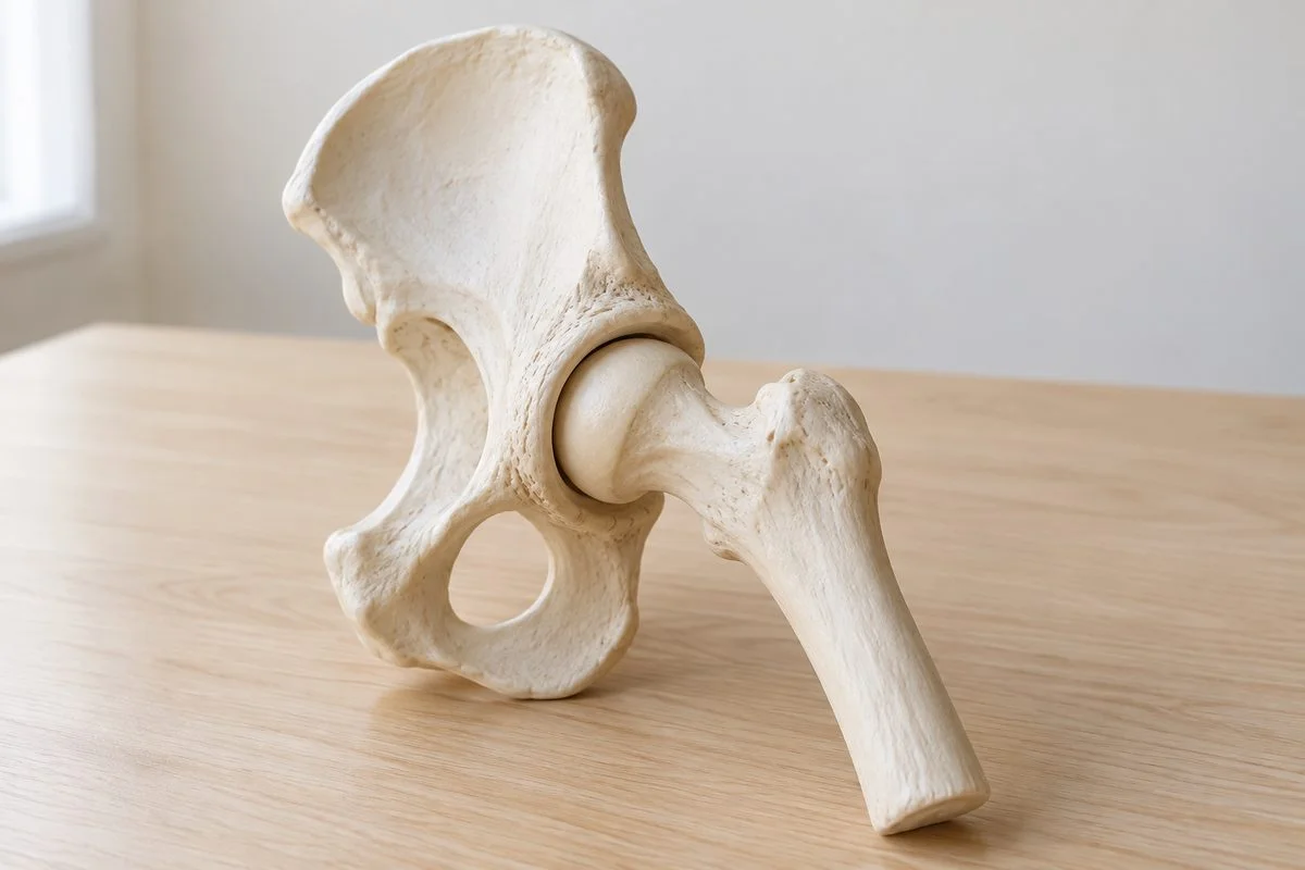
The hip's ball-and-socket joint allows changes of direction while walking.
If your child says…“The hip works like a hinge.”
Ask
Would a hinge allow the turning movement available at your shoulder?
What’s true
The rounded thigh-bone head fits the hip socket, allowing movement in several directions.

When movement structures are affected

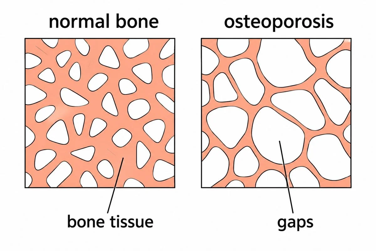
Two labelled bone networks show thinner connections and larger gaps in osteoporosis.

8. Disorders of muscular and skeletal systems

Disorders can affect bones, muscles, joints or nerve-to-muscle communication.

Show
Look at the two bone-network diagrams. Find the thicker connections on the left and larger gaps on the right.
Ask
Which patch would provide less support?
Say
The right patch contains less supporting bone, as in osteoporosis.
Used in
Recognising reduced bone mass helps explain why some bones fracture more easily.
If your child says…“Osteoporosis makes bones heavier and harder to break.”
Ask
Does the patch with wider gaps contain more bone or less?
What’s true
Osteoporosis reduces bone mass and increases the likelihood of fractures.

A real problem

An artificial hip relieved pain while preserving movement.

Charnley's successful hip implant entered use in 1962.

It paired an artificial thigh-bone head with a plastic socket.

The operation relieved hip pain while maintaining joint movement and mobility.

John Charnley TrustYale University LibraryWWL Teaching Hospitals

An anatomical hip-joint model on a classroom desk.

Concept Lab · Common misconception

The tempting mistake in Locomotion and Movement

Open standalone misconception page →

Do muscles push bones to move them?

Wrong

The biceps pushes the forearm up.

Why it feels right. The forearm moves in the direction I think of as up, so the muscle must push it.

Right

A contracting muscle pulls on its attachment. Opposing muscles can move a joint in opposite directions.

Worked example

Flexion
Biceps contracts→Tendon pulls→Elbow bends
Extension against resistance
Triceps contracts→Tendon pulls→Elbow straightens
Muscles pull on tendons. Opposite muscles move the elbow in opposite directions.
  1. Bend your elbow.
  2. The biceps shortens and pulls on its tendon.
  3. Straighten the elbow against resistance and the triceps pulls from the opposite side.

Ask your child eight quick questions.

One per step. The answer and the reason are given.

  1. A bird flaps while remaining on its perch. Which category describes this?

    Movement without locomotion. Its wings move, but the bird remains in the same place.

  2. Which two lines mark the ends of one complete sarcomere?

    Two neighbouring Z lines. The middle M line lies inside the unit.

  3. Which protein receives calcium's signal to expose actin's attachment sites?

    Troponin. Calcium binding changes the arrangement of troponin and tropomyosin.

  4. Which muscle band keeps its length during contraction?

    The A band. Sliding changes filament overlap, not the length of thick filaments.

  5. Which skeletal division contains the breastbone (sternum)?

    The axial skeleton. The sternum belongs to the central chest framework.

  6. Which skeletal division contains the patella?

    The appendicular skeleton. The kneecap belongs to a lower limb.

  7. Which joint shape allows the thigh to move in several directions?

    Ball-and-socket. A rounded head can turn within its socket.

  8. What happens to bone mass in osteoporosis?

    It decreases. Loss of supporting bone tissue makes fractures more likely.

15 more questions with answers, 4 with pictures